Spontaneous Resolution of a Patient with a Symptomatic Pheochromocytoma
Jun 19, 2024
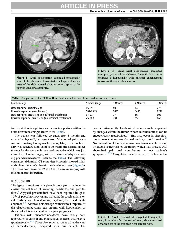
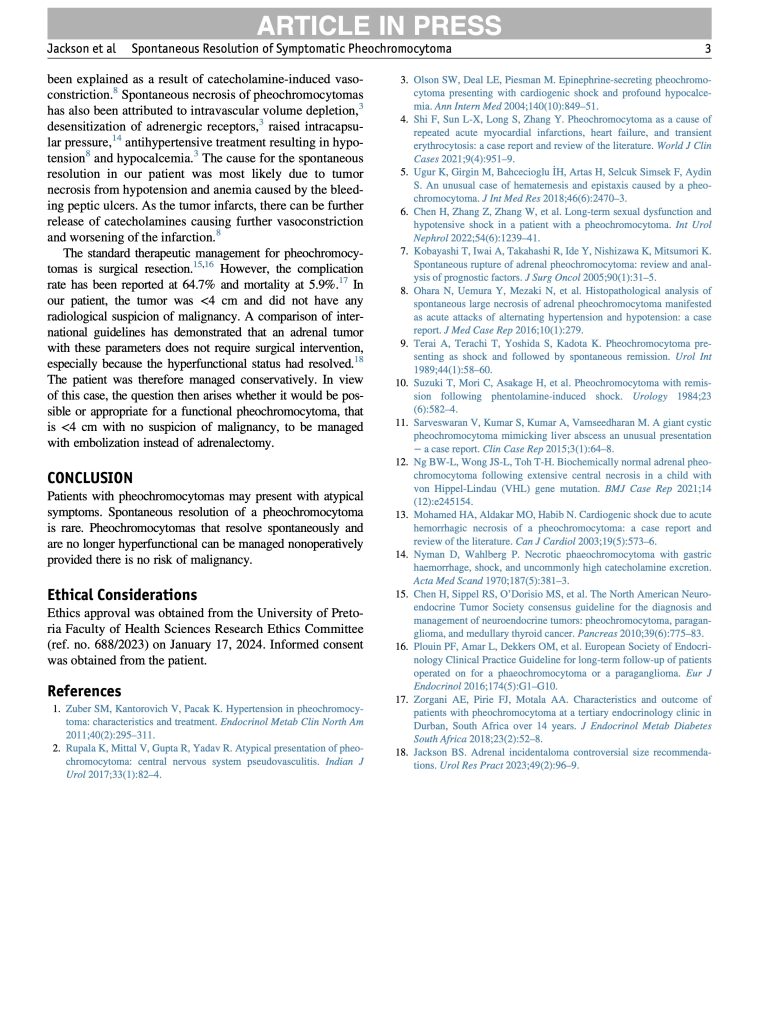
An article in press in The American Journal of Medicine describes an unusual clinical course in a 56-year-old woman with a symptomatic pheochromocytoma managed at Kalafong Provincial Tertiary Hospital in Pretoria. She presented with a two-year history of persistent right upper quadrant abdominal pain with intermittent nausea, vomiting, and unintended weight loss. Despite the classic association of pheochromocytoma with fluctuating hypertension, her blood pressure was stable and well controlled on hydrochlorothiazide and amlodipine. CT imaging revealed a 3.8 x 2.8 x 3.3 cm hyperenhancing retroperitoneal mass inseparable from the right adrenal gland, closely abutting the inferior vena cava without invasion. Biochemical testing confirmed the diagnosis, with elevated 24-hour urinary metanephrines and normetanephrines, and markedly raised vanillylmandelic acid markers, while multiple endocrine neoplasia type II was excluded. While awaiting adrenalectomy, she deteriorated with severe pain, hypotension, and profound anemia related to gastrointestinal bleeding from NSAID-associated gastric ulcers. Repeat contrasted CT then showed the adrenal mass had become hypodense with minimal enhancement, suggesting infarction and necrosis. The case highlights how endocrine tumors can present atypically and evolve unexpectedly, underscoring the value of careful imaging and biochemical follow up. It also reinforces the need for multidisciplinary care and caution with nonsteroidal anti-inflammatory self-medication.